Products
|
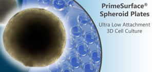
The inner wall of PrimeSurface products are coated with our unique ultra hydrophilic polymer, which prevents a cell from adhering to the plastic surface induces a spontaneous cell spheroid formation in equal size and shape. |
Features
- Ideal tool for uniform spheroid cell culture formation
- The uniform spheroid cell culture could be effective formed due to the adhesion of cell is inhibited by our unique surface coating technique of PrimeSurface product.
- Ideal tool for differentiation study
- Inducer reagent for differentiation can be applied after EB body formation of ES cells.
- The chemical bonding of ultra-hydrophilic coating polymer on our product is stable culture surface which makes no elution of chemicals from substrate.
- Ideal for differentiation and stimulation response studies
Applications
Medical Research Institution/Laboratory
Cell Culture
For Stem Cell Research:
Induction of ES, iPS and mesenchymal stem cells differentiation by embryoid body formation.
For Drug Screening Research & Development:
An ideal product for drug response studies. Three dimension spheroid models are more physiologically relevant than two dimension monolayer model.
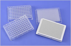
Specifications
| Cat. No | Product | Description | Culture Area | Volume | Qty/Pk | Qty/Cs |
|---|---|---|---|---|---|---|
| MS-9035XZ | PrimeSurface Dish 35 Φ | 35 Φ × 14(H)mm | 9 cm2 | - | 5 | 50 |
| MS-9060XZ | PrimeSurface Dish 60 Φ | 60 Φ × 15(H)mm | 21 cm2 | - | 10 | 100 |
| MS-9090XZ | PrimeSurface Dish 90 Φ | 90 Φ × 20(H)mm | 57 cm2 | - | 10 | 50 |
| MS-9024XZ | PrimeSurface Plate 24 | 24wells, Flat | 1.8 cm2 | 3.4mL/well | 1 | 10 |
| MS-9384UZ | PrimeSurface 384U | 384 | U bottom | 0.1mL | 1 | 20 |
| MS-9384WZ | PrimeSurface 384U White Plate | 384 | U bottom | 0.1mL | 1 | 20 |
| MS-9096VZ | PrimeSurface 96V | 96 | V bottom | 0.3mL | 1 | 20 |
| MS-9096MZ | PrimeSurface 96M | 96 | Spindle bottom | 0.2mL | 1 | 20 |
| MS-9096UZ | PrimeSurface 96U | 96 | U bottom | 0.3mL | 1 | 20 |
| MS-9096WZ | PrimeSurface 96U White Plate | 96 | U bottom | 0.3mL | 1 | 20 |
Remark
- Storage: Room temperature.
- Expiration: 2 years after production
Experiments
For Tissue engineering and Regenerative medicine:
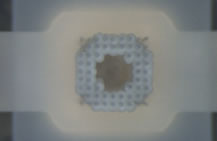
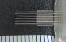
Regenova is a novel robotic system that facilitates the fabrication of three- dimensional cellular structures by placing cellular spheroids in fine needle arrays according to pre-designed 3D data. Followings are examples of such fabrications using S-BIO's PrimeSurface™ 96U plate and Bio 3D Printer, Regenova (Cyfuse Biomedical K.K.).; neural 3D tissue and 3D tissue with mesenchymal stem cells.
Neural 3D tissue
| Cell sources | : | hiPSC derived neural progenitor cells |
|---|---|---|
| Quantity of cells | : | 4 X 10⁴ cells/well |
| Culture Medium | : | For neural cells |
| Maturation duration on Well Plate | : | 2 days |
| 3D structure of printed 3D tissue | : | printed with 3 X 3 X 2 |
| Quantity of spheroids used for 3D tissue | : | 18 spheroids |
| Maturation duration after 3D printing | : | Remove needles 9 days after 3D print |

Spheroid 3D tissue after removal of needles
3D tissue with mesenchymal stem cells
| Cell sources | : | human adipose tissue derived mesenchymal stem cells (hADSC) |
|---|---|---|
| Quantity of cells | : | 5 X 10³ cells/well |
| Culture Medium | : | For MSC |
| Maturation duration on Well Plate | : | 2 days |
| 3D structure of printed 3D tissue | : | Circle shape with 48 spheroids x 10 layers |
| Quantity of spheroids used for 3D tissue | : | 480 spheroids |
| Maturation duration after 3D printing | : | Remove needles 6 days after 3D print |
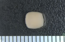
Spheroid After 3D print, from above After 3D print, side view 3D tissue after removal of needles
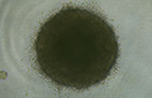
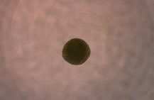
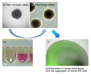
Application example of hES cells differentiation using PrimeSurface
The uniform spheroids form spontaneously within one day for effective aggregate formation of ES cells by using PrimeSurface
Self-formation of neural retina tissue from the aggregate of human ES cells by using PrimeSurface MS-9096V
| Culture plate | : | PrimeSurface MS-9096V |
|---|---|---|
| Cell type | : | Human ES cells (KhES-1 strain) |
| Seeding density | : | 9,000 cells/well |
| Culture medium | : | GMEM+KSR+NEAA+2ME+ 20uM Y-27632 |
| Culture environment | : | 5%CO2, 37°C |
Data resource
- Picture a-c
Division of Human Stem Cell Technology RIKEN Center for Developmental Biology
Reference
- Self-Formation of Optic Cups and Storable Stratified Neural Retina from Human ESCs; NakanoT, Ando S, Takata N, Kawada M, Muguruma K, Sekiguchi K, Saito K, Yonemura S, Eiraku M, Sasai Y; Cell Stem Cell, 10 (6), 771-785 (2012)
Evaluation example of anticancer drug efficacy by using PrimeSurface
| Cell type | : | MCF-7 (Human breast cancer cell) |
|---|---|---|
| Anticancer drug | : | 5-Fluorouracil (5-FU) |
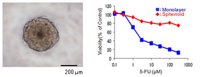
Data resource
Nishio Lab., Department of Genome Biology, Kinki University School of Medicine
Please check the supplemental data for more detail example of application.

Supplemental Data
Time-lapse imaging of human iPS cells spheroid (embryoid body) formation
[Culture conditions]
| Culture plate | : | PrimeSurface MS-9096V |
|---|---|---|
| Cells | : | hiPS cell (201B7: Takahashi K et al. Cell, 2007 Nov 30;131(5):861-72, iPS Academia Japan, Inc.) |
| Seeding density | : | 9,000 cells/well |
| Culture medium | : | DMEM/F12 + 20% (v/v) KSR + 1% (v/v) NEAA + L-Glutamine (2mM) + Β-Mercaptethanol (80μM) + Y-27632(30μM) |
| Culture environment | : | 5%CO2, 37°C |
Time-lapse imaging of HeLa cells spheroid formation in 96 well plate
[Culture conditions]
| Seeding density | : | 1,000 cells/well |
|---|---|---|
| Culture medium | : | MEM+10FBS 100μL/well |
| Culture period | : | 48hours |
Time-lapse imaging og HepG2 cells spheroid formation in 384 well plate
[Culture conditions]
| eding density | : | 1,000 cells/well |
|---|---|---|
| Culture medium | : | DMEM (Low Glucose) + 10%FBS 50μL/well |
| Culture period | : | 48hours |
PrimeSurface Product list